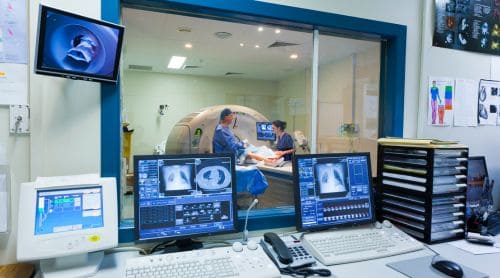
High-level summary: Interventional radiology (IR) is a minimally invasive specialty where radiologists use imaging guidance to diagnose and treat conditions that once required open surgery. Nurses play a critical role in patient preparation, moderate sedation monitoring, and post-procedure care. Understanding IR basics helps nurses prepare patients, anticipate complications, and deliver safer outcomes.

Radiology has evolved beyond mere x-rays. Over the past few decades, interventional radiology has transformed from a purely diagnostic service into one of medicine’s most dynamic treatment specialties. Procedures that once required surgery, general anesthesia, and days of recovery can now often be completed in a radiology suite, with the patient going home the same day.
For nurses, that evolution matters. Patients increasingly arrive on your unit having just come from IR, or they’re being prepared to go there. Knowing what happens in that room, and why, puts you in a much better position to educate patients, monitor for complications, and coordinate care effectively.
This post breaks down the essentials: what interventional radiology is, how the team is structured, how patients are selected and prepared, what common procedures involve, and what your role looks like before and after the procedure.
Related CE course for nurses: A Review of Interventional Radiology
What is interventional radiology, and how does it differ from diagnostic radiology?
Diagnostic radiology uses imaging—x-ray, CT, MRI, ultrasound, nuclear medicine—to identify what’s wrong. Interventional radiology takes the next step. It uses those same imaging tools to guide minimally invasive procedures that treat the problem. Think of it this way: diagnostic radiology answers the question, while interventional radiology acts on the answer.
According to the Society of Interventional Radiology (SIR), there are more than 8,000 interventional radiologists in the United States. The advantages of IR over traditional surgery include:
- Most procedures can be done in an outpatient setting
- General anesthesia is usually not required
- Risk, pain, and recovery time are significantly reduced
- Costs are often lower than surgical alternatives
IR suites function essentially as minor operating rooms. They’re equipped with fluoroscopy systems, hemodynamic and cardiac monitoring, emergency medications, and resuscitation equipment. Patients who might once have faced lengthy surgical hospitalizations can now be treated and discharged within hours.
Who’s on the interventional radiology team, and where does the nurse fit in?
The IR team includes the interventional radiologist, specially trained cardiovascular interventional technologists, and a dedicated IR nurse.
The radiologist performs the procedure. The technologists manage imaging equipment, contrast injectors, and technical positioning. The nurse’s primary role is patient care and monitoring throughout the procedure. That’s a critical distinction. The IR nurse isn’t just there to assist with the procedure. Their main job is to continuously assess the patient, monitor cardiac rhythms, administer medications, and respond to complications.
Because of this, IR nursing requires a critical care background. The American Radiological Nurses Association (ARNA), recognized by the American Nurses Association, offers a Certified Radiological Nurse (CRN) credential for nurses in this specialty. IR nurses are also expected to hold ACLS certification, and in some settings, PALS as well.
Even if you’re not the IR nurse, understanding this dynamic helps you know what your patient has been through and what the IR team was managing during the procedure.
How are patients selected for interventional radiology procedures?
Not every patient is a candidate for IR, even when a procedure seems straightforward. Careful pre-procedure assessment is essential.
What lab values matter before an IR procedure?
Before any invasive procedure, the following results should be reviewed:
- Platelet count: should not be below 50,000/mm³
- INR: generally should be less than 1.5 to 1.7
- PT and PTT: within normal limits
- BUN and creatinine: BUN greater than 23 mg/dL or creatinine greater than 1.2 mg/dL signals caution when contrast agents are involved
Patients on anticoagulants require specific management. Providers typically stop heparin four hours before a procedure. Warfarin (a vitamin K antagonist) may need to be held for four to seven days because of its longer half-life. Always confirm the plan with the ordering team.
Patients with renal insufficiency, congestive heart failure, multiple myeloma, or diabetes managed with metformin face additional risks when iodinated contrast is used. A thorough medical history, including herbal supplements and over-the-counter medications like aspirin, is essential.
What should nurses know about contrast agent reactions?
Most IR procedures use iodinated contrast agents to visualize blood vessels and organs. These agents can cause reactions ranging from mild (flushing, hives, nausea) to severe (anaphylactic shock, cardiac arrest).
Patients with a known history of contrast reactions may be premedicated. A common protocol includes methylprednisolone 32 mg given at 12 hours and 2 hours before the procedure, plus diphenhydramine 50 mg given one hour before. Always confirm the institutional protocol and ensure the patient has an IV in place prior to the procedure.
Gadolinium-based contrast agents (GBCAs), used in MRI, carry a separate risk: nephrogenic systemic fibrosis (NSF) in patients with severe renal insufficiency. The FDA has mandated boxed warnings on all nine GBCAs available in the US. If your patient has compromised kidney function and is heading for a contrast-enhanced study, flag it early.
How are patients prepared for an interventional radiology procedure?
Preparation starts well before the patient arrives in the IR suite.
Pre-procedure checklist for nurses
Nurses preparing patients for IR should confirm the following:
- NPO status: most patients should have nothing by mouth after midnight; confirm whether local anesthetic only or moderate sedation is planned
- IV access: a patent peripheral IV must be in place before the procedure
- Consent: informed consent must be signed and witnessed before sedation is given
- Lab work: current results should be available and reviewed
- Medications: document all current medications, last doses taken, and any diabetes management (especially insulin or metformin)
- Allergies: confirm contrast allergy history and ensure premedication has been given if indicated
- Transport companion: patients receiving moderate sedation must have someone to drive them home and should not be left alone overnight
For inpatients, make sure all recent chart data, including EKGs, consult notes, imaging, and lab values, is available in the room before the procedure begins.
What is moderate sedation, and what does the nurse need to know?
Moderate sedation is used in almost all IR procedures. Allowing for minimally depressed levels of consciousness, moderate sedation lets the patient maintain a patent airway and respond to commands without requiring general anesthesia.
The goals are:
- Anxiety reduction
- Pain control
- Partial amnesia
- Safe return to normal activity
Common moderate sedation medications used in IR
Opioids (for pain control):
- Fentanyl – 100 times more potent than morphine; dosed in micrograms
- Morphine and meperidine are also used
Sedatives/benzodiazepines (for anxiety and amnesia):
- Midazolam – the drug of choice at many facilities; 3–4 times more potent than diazepam, with shorter duration
- Diazepam and lorazepam are alternatives
Reversal agents to keep on hand:
- Naloxone: reverses opioid effects
- Flumazenil: reverses benzodiazepine effects
Respiratory depression is the primary adverse effect of IV moderate sedation. The Joint Commission requires continuous pulse oximetry for all patients receiving moderate sedation. Nurses must monitor cardiac rhythm, oxygen saturation, blood pressure, and level of consciousness throughout the procedure.
The Modified Aldrete Score is commonly used to determine when a patient is ready for discharge. A score of 18 or higher indicates readiness. Criteria include activity, respiration, circulation, consciousness, oxygen saturation, pain, ambulation, and urine output.
What procedures should nurses be familiar with?
IR covers a broad and expanding range of procedures. Here’s a practical overview of some of the most common ones:
Biopsies
IR uses CT, ultrasound, MRI, or fluoroscopic guidance to sample tissue from organs including the liver, kidney, lung, pancreas, and bone. The technique avoids the need for open surgery and allows same-day discharge in most cases. Complications vary by site but include bleeding and, for lung biopsies, pneumothorax, which occurs in approximately 9% to 54% of thoracic biopsies.
Percutaneous drainages
Fluid collections, abscesses, and cysts in the abdomen, pelvis, and chest can be drained using image-guided catheters. Patients with intra-abdominal abscesses who are already on antibiotics may still develop sepsis after drainage. Nurses caring for these patients post-procedure must watch closely for signs of infection and hemodynamic instability.
Percutaneous nephrostomy (PCN)
PCN provides access to the renal collecting system to relieve obstruction. Patients undergoing PCN must receive IV antibiotics within one hour of the procedure. Expect post-procedure hematuria for one to two days. Explicit tube care orders and output documentation are essential.
Angiography and angioplasty
Angiography uses contrast injected via catheter to image blood vessels. Balloon angioplasty (percutaneous coronary intervention, or PCI) opens narrowed vessels by inflating a balloon catheter at the point of stenosis. Patients may experience discomfort or a sensation of warmth or “urinating” when contrast is injected in the pelvic region. Be sure to warn them in advance to prevent distress.
Post-angioplasty, the puncture site (usually the femoral artery) requires close monitoring. The leg must be kept straight for several hours. Both extremities should be assessed, even if only one side was punctured. A cool or discolored leg opposite the puncture site may indicate a vascular complication.
Vascular stents and filters
Stents hold vessels open after angioplasty. Providers place IVC filters in the inferior vena cava to prevent pulmonary emboli in patients who can’t receive anticoagulants. According to the FDA, remove retrievable IVC filters as soon as protection from PE is no longer needed. Nurses should flag filters that have been in place for extended periods during hand-offs.
Embolization
Intentional embolization blocks blood flow to a specific area. It’s used to treat uterine fibroids, arteriovenous malformations, active hemorrhage, and tumor-related bleeding. Post-embolization syndrome, including pain, nausea, vomiting, and fever, is normal and typically self-limiting. Supportive care is the standard management approach.
Tumor ablation
Radiofrequency ablation (RFA) uses heat (60–100°C applied for 7–15 minutes on average) to destroy tumors in organs like the liver, kidney, and lung. Microwave ablation works similarly but at higher energy, which allows treatment of larger tumors. Post-procedure nausea, vomiting, fever, and pain are common. Serious complications include pneumothorax and hemoptysis.
TIPS (Transjugular Intrahepatic Portosystemic Shunt)
TIPS creates a shunt within the liver to reduce portal hypertension and control variceal bleeding. It’s a significant procedure that may require moderate to heavy sedation or general anesthesia. Providers usually discharge patients within two to four days. Hemorrhage and bile duct trauma are the major complications.
What is the nurse’s role in post-procedure care and patient education?
Once a patient returns from IR, your assessment should cover:
- Vital sign stability
- Return of reflexes and motor control
- Level of consciousness (should be at or near pre-procedure baseline)
- Puncture site integrity—look for swelling, bleeding, or hematoma
- Extremity assessment after angiographic procedures
- Urine output for patients who received contrast or had urologic procedures
What should nurses teach patients before discharge?
Make sure discharge education is both verbal and written. Key points include:
- No driving or operating machinery until the following day after moderate sedation
- No alcohol until the next day
- Who to call if symptoms worsen and what symptoms to watch for
- Site care for any drainage catheters or access points
- When to return for follow-up imaging or catheter changes
For patients going home with drainage catheters, education should include catheter care, output measurement, signs of infection, and emergency contact information. An interventional radiologist must remove any radiologically placed drainage catheter, not the referring physician.
Patients who’ve had lung biopsies should be warned to seek help immediately for sudden breathing difficulty (beyond expected site soreness). Patients with hepatic or pancreatic procedures need to know that fever, vomiting, chills, and increasing pain may signal a serious complication.
Radiation safety: What nurses should know about the ALARA principle
Medical radiation now represents the largest man-made source of ionizing radiation exposure. In interventional radiology specifically, CT and fluoroscopy deliver some of the highest radiation doses of any common imaging procedure.
The principle of ALARA (As Low As Reasonably Achievable) is a federal regulatory requirement for all radiation safety programs. As a nurse, you may not control imaging decisions, but you can advocate for nonionizing alternatives (like ultrasound or MRI) for follow-up imaging when clinically appropriate, and flag unnecessary repeat imaging during care coordination.
What do nurses need to know about interventional radiology?
The IR nurse role is critical care, not just procedural support. Nurses in IR must be prepared to manage hemodynamic changes, cardiac rhythms, medication administration, and emergency response, all during an active procedure.
Pre-procedure preparation is a shared responsibility. Ward nurses, office nurses, and IR nurses all contribute to safe outcomes by confirming lab values, managing anticoagulants, completing allergy histories, and preparing patients and families for what to expect.
Post-procedure monitoring requires attentiveness to subtle changes. A cool extremity, a swelling puncture site, or a slowly dropping blood pressure may be the first sign of a serious complication.
Patient education is non-negotiable. Many IR patients go home the same day with drainage catheters, healing puncture sites, or post-procedure syndrome in progress. Clear, reinforced discharge teaching directly impacts outcomes.
Interventional radiology is expanding rapidly. New procedures, devices, and imaging technologies emerge regularly. Staying current, whether through continuing education, peer collaboration, or formal IR nursing training, helps you deliver the kind of care that makes a real difference for patients navigating these complex, often life-changing procedures.
Frequently asked questions about interventional radiology for nurses
What is the primary difference between interventional radiology and surgery?
Interventional radiology uses imaging guidance to perform minimally invasive procedures through small punctures or incisions, rather than open surgical access. Most IR procedures don’t require general anesthesia, carry lower complication rates, and involve significantly shorter recovery times than equivalent surgical approaches.
What lab values does a nurse need to verify before an IR procedure?
Key labs include platelet count (should be above 50,000/mm³), INR (generally below 1.5–1.7), PT and PTT, and for contrast procedures, BUN and creatinine. Patients with elevated BUN (greater than 23 mg/dL) or creatinine (greater than 1.2 mg/dL) are at risk for contrast-induced nephropathy.
What medications are typically used for moderate sedation in IR?
The most common combination is a benzodiazepine (usually midazolam) for sedation and amnesia, plus an opioid (often fentanyl) for pain control. Reversal agents—naloxone for opioids and flumazenil for benzodiazepines—must be immediately available during any procedure using moderate sedation.
What is postembolization syndrome, and how should nurses manage it?
Postembolization syndrome is an expected response following embolization procedures. It includes pain, nausea, vomiting, and low-grade fever caused by the body’s reaction to blocked blood flow and tissue changes. The condition is self-limiting, and care is supportive: antiemetics, analgesics, hydration, and monitoring.
When is an IVC filter used, and what should nurses watch for after placement?
IVC filters are placed in patients at risk for pulmonary embolism who can’t receive anticoagulants. After placement, nurses should monitor for femoral or jugular puncture site complications, signs of deep vein thrombosis, and limb circulation. The FDA recommends removing retrievable filters as soon as PE protection is no longer needed. Nurses should document filter presence and alert the care team if removal hasn’t been addressed.
How should nurses prepare patients emotionally for interventional radiology procedures?
Anxiety is common, especially when patients have limited information. Nurses should explain what to expect using plain language, answer questions honestly, and emphasize that the radiology team will be monitoring them throughout. Patients receiving moderate sedation should understand they don’t need to be asleep—just comfortable and able to cooperate with brief instructions during the procedure.