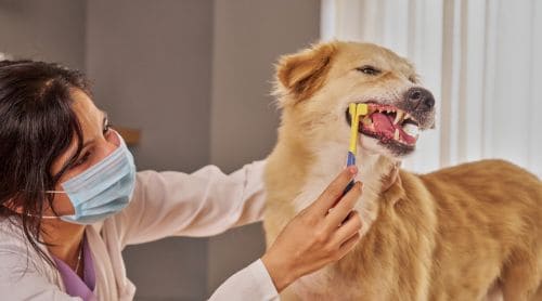
A comprehensive oral examination is the cornerstone of effective veterinary dentistry. It enables practitioners to detect early signs of disease, formulate appropriate treatment strategies, and educate clients on the significance of dental care. Performing a thorough oral examination requires more than a cursory inspection of the oral cavity. It needs a methodical, organized strategy and the appropriate instruments.
This article aims to assist small animal veterinarians, veterinary technicians, and students in enhancing their proficiency in doing thorough oral examinations. Whether you’re new to veterinary dentistry or looking to refine your current protocols, this guide provides systematic techniques to enhance patient outcomes and elevate care standards in veterinary dentistry.
Related CE course for veterinary professionals: Veterinary Dentistry: Basic Practical Guidebook of a Thorough Oral Examination, 2nd Edition

The oral examination
A comprehensive oral examination in veterinary practice is typically conducted in four distinct stages.
First stage: Obtaining medical history
The first stage involves obtaining a detailed history. Engage the owner with targeted questions and attentively listen to their observations. Ask dental-specific inquiries, such as changes in eating behavior, chewing preferences, drooling, halitosis, or evidence of oral pain, to gather a detailed patient history.
Owners may fail to recognize subtle symptoms; therefore, targeted inquiries can identify early indications of dental disease. A thorough medical and dental history can provide valuable context and may help clarify the nature and duration of the presenting oral condition(s).
Assessing the patient’s behavior and temperament is essential. Animals exhibiting anxiety, pain, or aggression may require sedation to ensure a safe and thorough examination. The safety of patients and staff should always be a priority.
Second stage: Extra-oral assessment
In the extraoral physical examination, the clinician visually inspects and palpates the head and neck of the conscious patient to detect abnormalities such as asymmetry, swelling, or pain. A systematic extraoral exam provides valuable clues that guide the rest of the oral assessment.
- Facial symmetry and swelling: Inspect and palpate the face for any asymmetry, swelling, or areas of pain. Abnormalities may suggest the presence of a dental abscess, neoplasia, or even a mandibular or maxillary fracture.
- Lymph nodes: Evaluate the submandibular lymph nodes bilaterally, noting their size, consistency, and symmetry. Enlarged or firm nodes may indicate localized infection, inflammation, or neoplastic changes associated with oral disease.
- Salivary glands: Palpate the salivary glands for signs of enlargement, tenderness, or firmness, which may point to sialadenitis or other salivary gland disorders.
- Temporomandibular joints (TMJ): Gently open and close the patient’s mouth, observing for reduced range of motion, discomfort, or crepitus. These findings can reflect trauma, arthritis, or temporomandibular dysfunction.
- Ocular and nasal discharge: Note the presence of ocular or nasal discharge, as this can be a secondary sign of oral-nasal fistulas, advanced periodontal disease, or other dental-related pathology.
Third stage: Intraoral examination
The next step involves a careful intraoral examination of the visible oral structures in the conscious patient. Begin by inspecting the lips and oral mucosa, noting any abnormalities such as masses, ulcerations, foreign bodies, or discoloration that may indicate underlying pathology. Examine the buccal mucosa for lesions, with particular attention to those that may result from malocclusion or dental trauma.
Next, examine the tongue, evaluating its symmetry, surface texture, and the presence of any foreign material, masses, or signs of injury. Using an appropriate light source, examine the hard and soft palates for structural defects, inflammatory changes, or proliferative lesions.
Perform a visual examination of the teeth and gums to identify any apparent calculus, gingivitis, absent teeth, or cracked teeth. This assessment is inherently limited by patient cooperation and discomfort. It serves as a fundamental basis for a more comprehensive evaluation under anesthesia.
Charting veterinary dentistry
Accurate dental charting is a cornerstone of veterinary dentistry, providing both a diagnostic reference and a legal medical record. One of the most essential components of this process is periodontal probing and charting, which enables the assessment and documentation of periodontal attachment loss, the hallmark of periodontitis.
Because periodontitis involves the destruction of the periodontium, including the periodontal ligament and alveolar bone, its extent can only be evaluated through a combination of probing and radiographic imaging.
Periodontal probing
Periodontal probing is performed using a blunt-ended periodontal probe. This probe measures the depth of the gingival sulcus or periodontal pocket. In dogs, a normal sulcus depth is less than 3 mm, while in cats, it is typically less than 1 mm. The probe should be held using a modified pen grip with a stable finger rest and gently “walked” along the sulcus, parallel to the long axis of each tooth, using light pressure.
In the presence of gingival recession, the probe can also be used to measure the distance from the cementoenamel junction (CEJ) to the free gingival margin. Attachment loss (AL) is calculated by adding the probing depth to the amount of gingival recession. Bleeding on probing (BOP) should also be recorded, as it is an early indicator of active periodontal inflammation.
In addition to the periodontal probe, a dental explorer is used to detect pulpal exposures, external resorptive lesions, furcation defects, caries, and residual calculus following debridement. A dental mirror may help visualize the palatal and lingual surfaces of teeth, especially in smaller patients.
All examination findings should be thoroughly documented using a dental chart. Accurate and detailed charting not only facilitates the development of a comprehensive treatment plan but also serves as a vital reference point for monitoring disease progression or treatment outcomes over time.
Veterinary dentistry indices
Common indices used in veterinary dentistry qualitatively grade the degree of whole-mouth calculus (Table 1) and gingivitis (Table 2) on ordinal scales of 0–3.
Table 1. Calculus grading
| Grade | Description/Characteristics |
| 0 (None) | No supragingival calculus |
| 1 (Mild) | Minimal supragingival calculus |
| 2 (Moderate) | Moderate supragingival calculus with subgingival deposition |
| 3 (Severe) | Heavy supragingival calculus with substantial subgingival deposition |
Table 2. Gingivitis grading
| Grade | Description/Characteristics |
| 0 (None) | Normal gingiva |
| 1 (Mild) | Mild swelling and slightly increased red color |
| 2 (Moderate) | Moderate swelling and redness, with bleeding upon periodontal probing |
| 3 (Severe) | Severe swelling and redness, with spontaneous bleeding |
Table 3. Stages of periodontal disease
| Stage | Gingivitis | Attachment Loss | Description |
| 0 | No | No | Clinically normal |
| 1 | Yes | No | Gingivitis only |
| 2 | Yes | < 25% | Early periodontal disease |
| 3 | Yes | 25%–50% | Moderate periodontal disease |
| 4 | Yes | > 50% | Advanced periodontal disease |
Fourth stage: Comprehensive extraoral and intraoral assessment
The fourth stage, conducted under general anesthesia, allows for a more comprehensive extraoral and intraoral assessment. This includes detailed inspection, dental probing, and diagnostic imaging, facilitating the identification of conditions that may not be apparent during conscious examination.
The anesthetized oral examination enables a thorough evaluation of not only the supporting structures of the teeth but also the surrounding soft tissues of the oral cavity, including the tongue, tonsils, hard and soft palates, and buccal mucosa.
Each stage builds upon the previous, contributing to a complete and accurate diagnosis while ensuring both patient safety and diagnostic precision.
Dental radiography
Intraoral dental radiography, in particular, is indispensable in modern veterinary dentistry. It offers clinicians a detailed assessment of anatomical structures that are not visible upon routine examination. These high-resolution images provide a clear visualization of the teeth, pulp cavity, periodontal ligament space, alveolar bone, and surrounding structures. This level of detail is essential for diagnosing a wide range of conditions, such as periodontal disease, endodontic pathology, tooth resorption, and unerupted or impacted teeth.
Incorporating dental radiographs into both diagnostic and treatment protocols significantly enhances diagnostic accuracy, guides clinical decision-making, and ultimately improves patient outcomes.
Indications for dental radiography
- Assessment of periodontal disease, particularly to evaluate the extent of alveolar bone loss.
- Identification of missing or unerupted teeth that are not visible upon clinical inspection.
- Detection of tooth resorption, a condition commonly seen in feline patients.
- Evaluation of root structure and integrity prior to dental extractions, ensuring safe and effective removal.
- Investigation of periapical pathology, especially in cases following endodontic procedures or where there is suspicion of chronic infection.
- Diagnosis of oral trauma, such as mandibular or maxillary fractures.
- Detection of soft tissue abnormalities, including neoplasia, cysts, or other masses affecting the oral cavity.
Dental examination tools
A comprehensive oral examination requires a specific set of tools to be both effective and safe. Before proceeding, make sure your dental examination tools are ready. A basic dental examination will need:
- Veterinary headlamp or other light source with cold beam
- Dental mirror
- Periodontal probe
- Dental explorer
- Forceps
- Gauze sponges
- Protective eyewear with or without magnification
- Disclosing solution to detect plaque
- Digital camera with macro capability
- Personal protective equipment (PPE) e.g. gloves, masks, etc.
For a thorough dental examination under anesthesia, you will need:
- Dental radiography unit
- Anesthesia machine
- Appropriate patient monitor
Conclusion
Thorough, routine oral examinations are the cornerstone of veterinary dental care. They allow for the early detection of pathology, preventing pain, infection, and systemic complications. By combining a systematic approach with diagnostic imaging and accurate charting, veterinarians can provide a higher standard of care.
Beyond the clinical benefits, proactive dental assessments also enhance client satisfaction and compliance, strengthening long-term relationships. As the field of veterinary dentistry advances, continued education is essential to ensure that practitioners remain current with best practices, diagnostic tools, and treatment modalities. Investing in dental training elevates a practitioner’s skills and improves the overall quality of care for all patients.